Your Account
2024 © D'vakaso.
Designed by Zeptt Technologies
Designed by Zeptt Technologies
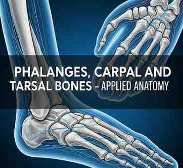
Bones of the hand and foot are essential for grasping, locomotion, and stability.
These include phalanges (finger and toe bones), carpal bones (wrist bones), and tarsal bones (ankle bones).
These are part of the appendicular skeleton and are studied in Rachana Shareera under Asthishareera.
PHALANGES
DESCRIPTION
Phalanges are the digital bones present in both the hands (fingers) and feet (toes).
Each finger has 3 phalanges (proximal, middle, distal) except the thumb/big toe which has 2 phalanges.
Total number:
Hand: 14 phalanges (5 digits × 3 each, thumb has 2)
Foot: 14 phalanges (similar arrangement as hand)
Each phalanx has:
Base (proximal)
Shaft
Head (distal)
SANSKRIT REFERENCE
अङ्गुल्यः पञ्च संख्याताः प्रत्येकस्य त्रयोऽस्थयः।
अङ्गुष्ठस्य तु द्वे प्रोक्ते चरकः सन्निबोधयत्॥
APPLIED ANATOMY
Fractures of phalanges are common due to crush injuries.
Dislocations at interphalangeal joints can limit movement.
Congenital anomalies: syndactyly (fusion), polydactyly (extra digits).
CARPAL BONES
DESCRIPTION
Carpal bones form the wrist joint and consist of 8 small bones arranged in 2 rows:
Proximal row (lateral to medial): Scaphoid, Lunate, Triquetrum, Pisiform
Distal row (lateral to medial): Trapezium, Trapezoid, Capitate, Hamate
These bones articulate with the radius, ulna, and metacarpals.
MNEMONIC (for identification)
Some Lovers Try Positions
That They Can’t Handle
SANSKRIT REFERENCE
मणिबन्धे अष्ट चास्थीनि स्थितानि स्युर्द्विधा युगे।
स्वल्पानि तानि कर्माणि हस्तस्य कुर्वते शुभम्॥
APPLIED ANATOMY
Scaphoid fracture is common in falls on an outstretched hand; risk of avascular necrosis due to poor blood supply.
Carpal tunnel syndrome: Compression of median nerve under flexor retinaculum, causing pain and numbness in hand.
Osteoarthritis can affect the carpometacarpal joint, especially of the thumb (first CMC joint).
TARSAL BONES
DESCRIPTION
Tarsal bones form the ankle and proximal part of the foot.
Total 7 tarsal bones:
Proximal group: Talus, Calcaneus
Intermediate bone: Navicular
Distal group: Cuboid, 3 Cuneiforms (medial, intermediate, lateral)
ARRANGEMENT
Talus articulates with tibia and fibula at ankle joint.
Calcaneus forms the heel and is the largest tarsal bone.
The arches of foot are supported by these bones for shock absorption and weight distribution.
SANSKRIT REFERENCE
गुल्फे च सप्त सम्पूर्णाः तालुकश्च अङ्घ्रिभूमिषु।
पृष्ठतो चलनायैव कुर्वन्ति दृढनिश्चयम्॥
APPLIED ANATOMY
Calcaneal fractures occur from height falls; can affect subtalar joint.
Flat foot (pes planus) arises due to collapse of medial longitudinal arch.
Talus is prone to avascular necrosis in fractures due to retrograde blood supply.
Tarsal tunnel syndrome: compression of posterior tibial nerve under flexor retinaculum.