Your Account
2024 © D'vakaso.
Designed by Zeptt Technologies
Designed by Zeptt Technologies
Patella is the largest sesamoid bone in the human body.
It is situated within the tendon of quadriceps femoris in front of the knee joint.
It plays a crucial role in knee extension and joint mechanics.
SANSKRIT REFERENCE
In Ayurvedic texts, the bones of the body are classified under अस्थिस्थूलानि and the region of the knee is referred as जानु प्रदेश.
The Patella may be correlated with जानुककुद् अस्थि mentioned in सुश्रुत संहिता.
सुश्रुतसंहिता शारीरस्थान ५/४७
यदस्ति जानुनो मध्ये स्थूलं च तद्विशेषतः।
जानुककुदिति ख्यातं स्थूलं तदस्त्यसंश्रयम्॥
FEATURES OF PATELLA
Type of Bone: Sesamoid bone (develops within a tendon).
Shape: Triangular, with apex directed downwards.
Location: Anterior to the knee joint, embedded in the quadriceps tendon.
Articulations: Articulates with the femoral condyles (patellar surface of femur).
Surfaces:
Anterior Surface: Rough, subcutaneous, covered by fascia and skin.
Posterior Surface: Smooth, articulates with the femoral condyles, covered by articular cartilage.
Borders:
Superior border (base): Attached to quadriceps tendon.
Inferior border (apex): Attached to patellar ligament.
Medial and lateral borders: Provide attachment to retinacula.
OSSIFICATION
Ossification begins from a single center during the 3rd to 5th year of life.
Complete ossification by puberty.
ATTACHMENTS
Tendons and Ligaments:
Superiorly: Quadriceps femoris tendon.
Inferiorly: Patellar ligament (continuation of quadriceps tendon).
Retinacula:
Medial and lateral patellar retinacula maintain patellar alignment and stability.
FUNCTIONS OF PATELLA
Increases the leverage of quadriceps muscle.
Protects the anterior aspect of the knee joint.
Prevents friction of quadriceps tendon over the femoral condyles.
Centralizes the pull of quadriceps femoris on the tibia.
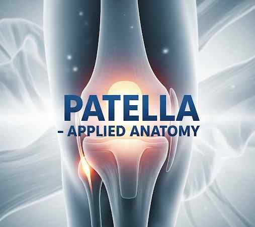
APPLIED ANATOMY
Patellar Fracture: Caused by direct blow or sudden contraction of quadriceps.
Patellar Dislocation: Usually lateral dislocation; common in young females due to a wider pelvis and increased Q-angle.
Chondromalacia Patellae: Softening and degeneration of cartilage under patella; common in athletes.
Patellectomy: Surgical removal of patella; results in weakened knee extension.
Jumper’s Knee: Inflammation of patellar tendon (Patellar tendinitis).
Prepatellar Bursitis (Housemaid’s knee): Inflammation of the bursa anterior to patella due to prolonged kneeling.
CLINICAL EXAMINATION AND DEMONSTRATION
Surface Marking: Patella is easily palpable and visible on anterior aspect of knee joint.
Mobility Test: In extension, patella is freely movable transversely. In flexion, it becomes fixed in the intercondylar groove.
Reflex Testing: Patellar tendon reflex (L2-L4 nerve roots) tested by tapping the patellar ligament.
MODERN CORRELATION (AS PER BD CHAURASIA'S HUMAN ANATOMY)
Volume 2 (Lower Limb):
Patella is described in detail under the chapter “Knee Joint”.
Details include: morphology, ossification, attachments, functions, and clinical anatomy.
Figures and Diagrams: Demonstrate patellar surface anatomy, relation with femur, and muscular attachments.
Clinical Notes:
Emphasize conditions like dislocation, congenital anomalies (bipartite patella), and fractures.
Orthopedic surgical relevance in knee replacements and trauma cases.